


Airway Management: Guidelines for Emergency Medicine
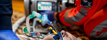
Airway management is one of the core competencies in emergency medicine. Neither effective ventilation nor adequate gas exchange can be guaranteed without open, secured airways. Evidence-based methods informed by current guidelines are used to safeguard pulmonary oxygenation and reduce the incidence of hypoxia-associated complications, increased mortality, and potential secondary harm.
These guidelines outline the definition, the key guidelines, and the central actions required for airway management – with a particular focus on prehospital use by the emergency medical services.
Airway management: the key facts in brief
- Airway management includes all the actions required to ensure airways are clear and ventilation is adequate – from positioning techniques to bag-valve-mask ventilation to endotracheal intubation.
- The current S1 Guidelines issued by the DGAI (Deutsche Gesellschaft für Anästhesiologie und Intensivmedizin [German Society for Anesthesiology and Intensive Care]) in 2023 recommend video laryngoscopy as the primary treatment for critically ill patients at risk of aspiration.
- Continuous capnography is obligatory after any form of airway management - in accordance with both S1-Leitlinie Prähospitales Atemwegsmanagement [S1 guidelines on prehospital airway management] and the 2021 ERC guidelines.
- The procedures range from non-invasive methods (manual maneuvers, mask ventilation, NIV) to invasive airway management (endotracheal tube, supraglottic devices) and are selected depending on clinical situation and experience.
- The endotracheal tube is considered the gold standard in invasive airway management.
What is airway management?
Airway management covers all the actions aimed at ensuring that the airways are clear and adequate spontaneous breathing or external ventilation are safeguarded.1
The primary aim is to maintain ventilation and an optimal oxygen supply, especially if spontaneous breathing is disrupted. This is frequently the case in emergencies, if a patient has suffered serious injuries, or during perioperative phases.
Indications for airway management
Airway management plays a central role in emergency medicine, in emergency medical services, in intensive care medicine and in anesthesia, as it forms the basis for adequate oxygenation and ventilation.
In a prehospital setting, effective airway management is often critical for a patient’s survival in resuscitation, emergency anesthesia or severe trauma cases.
The most important indications include:
Respiratory insufficiency
In respiratory insufficiency, the lungs are unable to guarantee an adequate oxygen supply to the body (hypoxemia) or effectively rid the body of excessive carbon dioxide (hypercapnia).
Danger of aspiration and risk of airway obstruction
For patients with an increased risk of aspiration, for example due to food, fluids or vomit, there is an acute danger of airway obstruction. Airway management prevents airways from being blocked by foreign bodies or aspirated material, thereby minimizing the risk of serious pulmonary complications such as aspiration pneumonia.
Traumatic injury to the head and upper airways
Trauma in the head and neck area, such as fractures of the facial skull or swelling of the upper airways, can significantly impair breathing.
Standards & guidelines for airway management
Airway management is structured by evidence and consensus-based guidelines which address treatment in both intra-hospital and prehospital settings. They define uniform standards for pre-anesthesiological evaluation, selection of method, check on position, and procedure in the event of a difficult airway and thus form a basis for safe, standardized, reproducible airway management.
The S1 guidelines are particularly relevant to emergency medicine in the German-speaking region.
S1 Guidelines on Airway Management 2023
The update to the S1 Airway Management Guidelines published in August 2023 (AWMF register number 001-028) was drafted under the chairmanship of the Deutsche Gesellschaft für Anästhesiologie und Intensivmedizin ([German Society for Anesthesiology and Intensive Care] - DGAI).1,2 It serves as a decision-making tool for medical personnel in anesthesia, intensive care medicine, and emergency medicine and includes over 100 numbered recommendations throughout the entire airway management process.1
Prehospital airway management: special features in the emergency medical services
For prehospital applications, there is a dedicated supplementary DGAI guideline to the S1 Guidelines on Prehospital Airway Management (AWMF 001-040).3 After all, prehospital airway management differs in many ways from the intra-hospital process: it is performed under pressure of time, frequently in poor light conditions, in cramped spaces, and under unforeseeable ambient conditions such as weather, noise or unfavorable positioning options.
The S1 guidelines of the DGAI, last thoroughly revised in 2019, formulate ten central recommendations and contain a structured algorithm for airway management in the emergency medical services.3,4
Its key recommendations include:
- Endotracheal intubation remains the gold standard in invasive airway management, although only for users with evidence of experience (at least 100 recorded intubations plus annual refreshers of at least ten intubations).
- Extraglottic airway management is the primary recommendation for those with inadequate experience of intubation.
- Bag-valve-mask ventilation should be performed using the “double-C” grip, optimal positioning of the head, a Guedel tube (oropharyngeal airway - OPA), and a high inspiratory oxygen fraction (FiO₂).
- Continuous capnography is obligatory after every airway management measure.
Airway management methods
A variety of measures are used in airway management to secure the airways and maintain unobstructed breathing. These range from basic manual techniques to invasive procedures and are selected on the basis of clinical situation and urgency.
Non-invasive methods
With non-invasive methods, there is no direct intervention in the airways.
Manual maneuvers
One of the frequently-used techniques is the cross-grip, which sees the thumb and index or middle finger used to open the oral cavity and to clear the airways.
The HTCL (head tilt and chin lift) maneuver is used on unconscious patients where spinal injury is not suspected. It involves tilting the individual’s head backwards to open the airway.
A further technique is the jaw thrust maneuver which involves stabilizing the head by means of specific movements - a particularly valuable measure in emergency situations requiring rapid action.
Stable position on one side
The stable position on one side is used as a positioning technique when artificial ventilation is impossible or not required. It stops patients aspirating vomit or fluids and ensures that their airways are temporarily clear.
Airway suctioning
If airways are obstructed by secretions, blood or foreign bodies, airway suctioning is required. The emergency medical services use portable, battery-operated suction devices for this; these ensure rapid, effective removal of secretions even under unpredictable conditions.
Mask ventilation
A classic example of this is manual bag-valve-mask (BVM) ventilation, where a ventilation bag with a ventilation mask is used to insufflate the lungs with oxygen by compressing the bag. This technique is especially suitable where an airway has not been secured or if the aim is to avoid an invasive procedure. It is used primarily when the airway does not need securing in the long term.
However, one study showed that patients treated with BVM had lower ROSC and survival rates compared to those whose airways were managed using an endotracheal tube and supraglottic devices.5 In addition, the effectiveness of bag-valve-mask ventilation has limitations in the case of prolonged transport times or serious airway diseases: in the case of prolonged transport times, it achieved less successful results in relation to oxygenation and ventilation than endotracheal intubation (ETI).6
Non-invasive ventilators
In addition to manual bag-valve-mask ventilation, other non-invasive ventilators are used to support breathing without invasive procedures. Non-invasive ventilation (NIV) has become established as an effective, gentle alternative to invasive airway management in numerous clinical scenarios.
In patients with acute respiratory insufficiency, in particular, NIV may reduce the requirement for endotracheal intubation and thus the risk of intubation-related complications. Furthermore, in certain patients groups, NIV improves survival rate and reduces the amount of time spent in intensive care units.7
Invasive airway management
In invasive airway management, airway devices are used to secure the airway directly. Invasive ventilation is essential in the case of severe respiratory arrest or inadequate manual ventilation.
Endotracheal tube
The endotracheal tube is considered the gold standard in invasive airway management, and involves inserting an endotracheal tube directly into the patient’s trachea via their mouth or nose.1,3 At the distal end is a cuff which protects against aspiration.
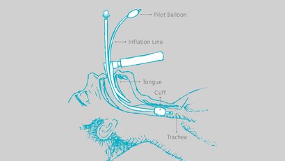
Studies show that the endotracheal tube achieves better results in terms of oxygen supply and ventilation compared to other methods, especially in the case of prolonged transport times.6 It is also linked to higher ROSC rates and a better survival rate during resuscitation.5
However, intubation does involve risks: success rates in studies vary widely (51–98%), so results need interpreting carefully. One comparative study showed no significant difference in neurologically favorable survival between endotracheal intubation and bag-valve-mask ventilation alone.8
Video laryngoscopy
In recent years, video laryngoscopy has moved from being a back-up technique to being the recommended method in the current guidelines.1,2 It involves using a laryngoscope blade with an integrated camera, the image being shown on an external display. A clear view of the glottis is thus obtained without precisely aligning the axes of mouth, pharynx, and larynx.
Evidence of the superiority of video laryngoscopy is consistent: the updated Cochrane Review by Hansel et al. (2022) showed that video laryngoscopes reduce the rate of failed intubation operations, improve the view of the glottis, and reduce the risk of laryngeal injuries, especially in patients with difficult airways.9
The DEVICE study exhibited significantly higher first-time intubation success rates under video laryngoscopy compared to direct laryngoscopy: 85.1% vs. 70.8%.10
Supraglottic and extraglottic airway management
If endotracheal intubation is difficult or not feasible from a time perspective, supraglottic or extraglottic airway devices can be a useful alternative. These devices are placed above the trachea to keep the airway open. They are quicker to position and require less experience than an endotracheal tube, though they do not provide reliable protection against gastric insufflation and aspiration, particularly in the context of emergency ventilation. In S1 Guidelines for Prehospital Airway Management, they are therefore recommended as a primary option only where there is limited experience of intubation.3,4
Common supraglottic/extraglottic airway management devices include:
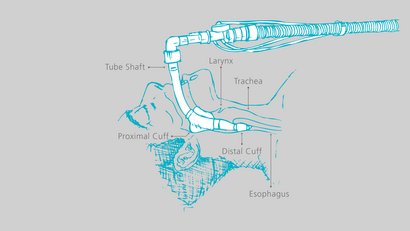
Laryngeal tube
The laryngeal tube has two cuffs. A large cuff (proximal cuff) seals the nasopharyngeal cavity, whilst a smaller one (the distal cuff) seals the entrance to the esophagus.
Standard laryngeal mask airway (LMA)
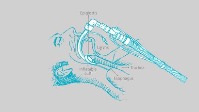
The laryngeal mask airway is a mask with an inflatable oval or teardrop-shaped attachment on the distal end and a valve to regulate the cuff. It is positioned in front of the larynx.
i-gel airway
This advanced version of the laryngeal mask airway has a non-inflatable seal whose gel-like texture allows it to fit over the larynx and minimize compression/displacement trauma.
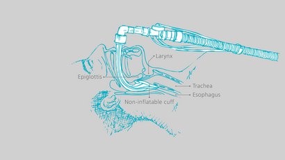
The laryngeal tube, LMA, and i-gel airway can be used as alternative emergency airways. One study showed that supraglottic airway management achieves a higher ROSC rate in resuscitation patients than pure bag-valve-mask ventilation.11
Compared to endotracheal intubation, however, it exhibits fewer positive effects on long-term survival and also a less favorable neurological outcome.8 Ventilation with supraglottic devices is also not ideal in every emergency situation which may impair the supply of air in complex positions.6
Hybrid strategy
If direct intubation is not possible at first, supraglottic airway management (e.g. laryngeal mask airway, laryngeal tube) is used as a temporary measure to gain time and create the conditions for intubation. As soon as the situation is stabilized, intubation is switched to the endotracheal tube for long-term airway management and optimal ventilation.
Coniotomy/tracheostomy
If conventional measures fail, performing a coniotomy as an emergency procedure or a tracheostomy as a surgical procedure may save the patient’s life. These procedures provide direct access to the trachea, but are risky and require highly qualified personnel.
Coniotomy, in particular, is considered a last-resort measure in a “Cannot Intubate, Cannot Oxygenate” (CICO) situation, when neither mask ventilation, supraglottic airway management nor endotracheal intubation can guarantee sufficient oxygenation.1,12
Measures following airway management
Verification of the correct position of the airway management device and continuous monitoring of ventilation are obligatory following any airway management measure. Capnography is specified as binding in both S1 Guidelines on Airway Management 20231 and in S1 Guidelines on Prehospital Airway Management3, as well as in the 2021 ERC Guidelines.13
Managing difficult airways
An airway is described as difficult when complications occur during airway management which render the use of standardized techniques such as mask ventilation or endotracheal intubation considerably more difficult or impossible.
A distinction is made here between the anticipated difficult airway, in which predictors for more difficult intubation or mask ventilation are obvious before airway management commences and the unanticipated difficult airway which only manifests itself in the course of airway management.1
Both scenarios may be due to anatomical anomalies (e.g. macroglossia, retrognathia), pathological changes (e.g. tumors, edema, trauma) or functional limitations (e.g. reduced mouth opening, restricted cervical spine mobility).
Predictors and classification of difficult intubation
As far as possible, a difficult airway should be detected and classified early on to allow the suitable airway management strategy to be selected. Clinically-established scoring systems are available for this purpose.
Mallampati score (Mallampati et al. 1985)
Before the procedure, an assessment is made of whether soft gums, uvula, and palatine arches are visible with the mouth open.14 The four-stage modified form of this approach usual nowadays estimates how difficult direct laryngoscopy is likely to be.
Cormack-Lehane classification (Cormack & Lehane 1984)
Rates the laryngoscopic view of the glottis during direct laryngoscopy in four grades - from complete view of glottis (Grade I) to no view of glottis and epiglottis (Grade IV).15 Unlike the Mallampati score, this assessment is not performed until the laryngoscopy has actually commenced.
The following applies to both classifications: the higher the value, the greater the risk of a difficult airway.
Managing an anticipated difficult airway
An anticipated difficult airway is involved if pre-anesthesiological predictors for more difficult mask ventilation, laryngoscopy or intubation are identified. For this scenario, the S1 Guidelines on Airway Management 2023 pursue the principle that airway management must be planned proactively and, if possible, completed with spontaneous breathing maintained.1
The guidelines define a clear hierarchy of procedures:
- Local anesthesia: In the event of an anticipated difficult airway, a method involving local anesthetic should be considered first. A back-up airway management concept should be communicated within the team and the materials for this prepared.1
- Airway management maintaining spontaneous breathing: if a general anesthetic is unavoidable and there are predictors of difficult intubation, airway management should be implemented with spontaneous breathing maintained. The flexible intubation endoscope has top priority in this situation.1
Managing an unanticipated difficult airway
An unanticipated difficult airway only manifests itself during airway management.
Algorithm: Plan A to Plan D
The guidelines produced by the Difficult Airway Society (DAS) in 2015 provide a simplified algorithm which has become established throughout the world for unanticipated difficult intubation procedures; these specify a sequential procedure:12
- Plan A: Endotracheal intubation (ETI)
- Plan B: Ventilation with a supraglottic airway device
- Plan C: Ventilation with a face mask
- Plan D: Emergency airway management at the throat
If Plans A to C fail, this means there is a “Cannot Intubate, Cannot Oxygenate” (CICO) situation. In this situation, the emergency coniotomy (Plan D) is the last emergency measure available.12,16
The role of video laryngoscopy in the difficult airway
Video laryngoscopy is a key piece of auxiliary equipment for an unanticipated difficult airway. If an unanticipated difficult airway is encountered, then use of a video laryngoscope is recommended if there is no success with direct laryngoscopy. The Cochrane review of 2022 evidences a significant reduction in failed intubations9, so the S1 guidelines 2023 require a video laryngoscope to be available at every workstation requiring airway management – including the emergency medical services.1,17
Airway management with WEINMANN
WEINMANN has an integrated portfolio for airway management by the emergency medical services – from clearing the airways to visualizing the glottis to invasive and non-invasive ventilation with integrated capnography.
ACCUVAC Pro airway suctioning
The ACCUVAC Pro portable suction device enables the rapid and effective removal of secretions, blood or foreign bodies in adults and children. Four individually adjustable suction levels and intuitive operation allow the device to be used both in emergency vehicles and in unpredictable environments.
MEDUVISION – video laryngoscope for prehospital intubation
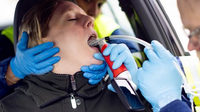
The MEDUVISION video laryngoscope was designed specifically for the requirements of the emergency medical services and supports implementation of the current guideline recommendation to prioritize video laryngoscopy in critically ill patients.1
Key features:
- Optimal view in any light conditions: the transflective allBRIGHT display can always be read, even in direct sunlight – a clear benefit outdoors and in brightly-lit areas of operation.
- Reliable control of tube position: the angle of the 3.2” portrait-format display can be flexibly adjusted to allow intubation with a clear field of view but with minimal reflection.
- Rugged for sessions: the display is dust-proof and protected against powerful jets of water (IP66), whilst the handle can withstand even long-term immersion (IP68). The device meets standard DIN EN 60601-1-12 for emergency medical treatment.
- Ready to use for an extended period: the high-performance battery can run for up to four hours and barely discharges when not in use - the device is ready to use even if it is used only rarely.
- Simple and safe to prepare: display and handle are easy to disconnect from one another, making setup easier. The disposable blades (Mac 2, Mac 3, Mac 4, HyMac 3) are directly interchangeable.
- Sustainable design: Rechargeable lithium battery and disposable blades made of upcycled material reduce consumption of resources.
This is how MEDUVISION addresses the specific requirements of prehospital airway management: a clear view of the glottis under difficult conditions, rapid readiness for operation and reliable function even when not used for extended periods.
MEDUVISION in use
MEDUMAT Standard² – ventilation with integrated capnography
The MEDUMAT Standard² ventilator delivers versatile solutions for invasive and non-invasive ventilation. Integrated RSI (Rapid Sequence Induction) mode supports controlled ventilation during drug-based anesthesia induction. Manual ventilation using MEDUtrigger supports combination with a variety of airway devices. Optional integrated capnography allows immediate and continuous checking on the position of the endotracheal tube in line with the recommendation in the guidelines.1,3,13
MEDUVENT Standard – ventilation without an external compressed gas supply
In extreme situations, the turbine-driven MEDUVENT Standard ventilator allows up to 7.5 hours of independent ventilation without an external supply of compressed gas, making it suitable for use in remote areas or under logistically demanding conditions. Manual mode using MEDUtrigger supports the use of a wide variety of airway devices.
Do you have any questions? Contact us.

Customer Service (International)
- Phone:+49 40 88 18 96 - 0
- Email:Please enable JavaScript to render this link!
Questions frequently asked about airway management
Airway management is the umbrella term for all the measures taken to assess, keep open, and secure the airways and to maintain sufficient ventilation and oxygenation - including position, suction, mask ventilation, securing with equipment, and post-procedure position check.
Securing the airway, on the other hand, describes the narrower part-step in which the airway is protected from displacement and aspiration by means of auxiliary equipment (e.g. endotracheal tube, laryngeal tube, laryngeal mask).
The first step is to inspect and assess the airways and to determine the urgency of securing the airways.
In the emergency medical services, the airway is secured under pressure of time, with limited light, in cramped conditions, and in an unforeseeable environment. The S1 Guideline for Prehospital Airway Management (AWMF 001-040) takes account of these particular features.3
1 AWMF-Leitlinienregister. S1-Leitlinie Atemwegsmanagement (AWMF-Registernummer 001-028, Stand 21.08.2023). https://register.awmf.org/de/leitlinien/detail/001-028
2 Piepho T, Kriege M, Byhahn C, et al. Wege zum sicheren Atemweg im Wandel: die S1-Leitlinie zum Atemwegsmanagement. AINS – Anästhesiologie · Intensivmedizin · Notfallmedizin · Schmerztherapie. 2024. https://www.thieme-connect.de/products/ejournals/html/10.1055/a-2421-6564
3 Timmermann A, Böttiger BW, Byhahn C, et al. S1-Leitlinie Prähospitales Atemwegsmanagement (AWMF-Registernummer 001-040, Stand 26.02.2019). https://www.awmf.org/leitlinien/detail/ll/001-040.html
4 Michael M, Bohn A, Hossfeld B, et al. Prähospitales Atemwegsmanagement. Notfallmedizin up2date / AINS, Thieme. 2020. DOI: 10.1055/a-1154-1959. https://www.thieme-connect.com/products/ejournals/pdf/10.1055/a-1154-1959.pdf
5 Tang Y, Sun J, Yu Z, et al. Outcome of Cardiopulmonary Resuscitation with Different Ventilation Modes in Adults: A Meta-Analysis. (2022).
6 Song M, Lee Y, Kim HJ, et al. Association Between Prehospital Airway Type and Oxygenation and Ventilation in Out-of-Hospital Cardiac Arrest. (2023).
7https://www.weinmann-emergency.com/de/themen/notfallbeatmung/nicht-invasive-beatmung
8Jung E, Kim HJ, Lee HJ, et al. Association of Prehospital Airway Management Technique with Survival Outcomes of Out-of-Hospital Cardiac Arrest Patients.
9 Hansel J, Rogers AM, Lewis SR, Cook TM, Smith AF. Videolaryngoscopy versus direct laryngoscopy for adults undergoing tracheal intubation. Cochrane Database of Systematic Reviews. 2022;4(4):CD011136. DOI: 10.1002/14651858.CD011136.pub3. https://pubmed.ncbi.nlm.nih.gov/35373840/
10 Prekker ME, Driver BE, Trent SA, et al. Video versus Direct Laryngoscopy for Tracheal Intubation of Critically Ill Adults (DEVICE-Trial). New England Journal of Medicine. 2023;389(5):418–429. DOI: 10.1056/NEJMoa2301601. https://www.nejm.org/doi/full/10.1056/NEJMoa2301601
11 Wang W, Zhang X, Liu M, Han X. Comparing Effectiveness of Initial Airway Interventions for Out-of-Hospital Cardiac Arrest: A Systematic Review and Network Meta-analysis of Clinical Controlled Trials.
12 Frerk C, Mitchell VS, McNarry AF, et al. Difficult Airway Society 2015 guidelines for management of unanticipated difficult intubation in adults. British Journal of Anaesthesia. 2015;115(6):827–848. DOI: 10.1093/bja/aev371. https://pmc.ncbi.nlm.nih.gov/articles/PMC4650961/
13 Soar J, Böttiger BW, Carli P, et al. European Resuscitation Council Guidelines 2021: Adult advanced life support. Resuscitation 2021;161:115–151. https://www.erc.edu/assets/documents/RESUS-8903-ALS.pdf
14 Mallampati SR, Gatt SP, Gugino LD, et al. A clinical sign to predict difficult tracheal intubation: a prospective study. Canadian Anaesthetists' Society Journal. 1985;32(4):429–434. https://link.springer.com/article/10.1007/BF03011357
15 Cormack RS, Lehane J. Difficult tracheal intubation in obstetrics. Anaesthesia. 1984;39(11):1105–1111. DOI: 10.1111/j.1365-2044.1984.tb08932.x. https://associationofanaesthetists-publications.onlinelibrary.wiley.com/doi/10.1111/j.1365-2044.1984.tb08932
16 Schlattmann L, Hartung B, Markwerth P. (Fehl-)Intubation im Rahmen der notfallmäßigen Koniotomie durch den Notarzt. Die Anaesthesiologie. 2026. DOI: 10.1007/s00101-026-01692 0. https://www.springermedizin.de/koniotomie/sicherung-der-atemwege/fehl-intubation-im-rahmen-der-notfallmaessigen-koniotomie-durch-/52444806
17 Deutsche Gesellschaft für Anästhesiologie und Intensivmedizin (DGAI). Update DGAI S1-Leitlinie "Atemwegsmanagement" (001-028) veröffentlicht. 2023. https://www.dgai.de/aktuelles-patientinnen-projekte/aktuelle-meldungen/aktuelle-meldungen-2023/1233-update-dgai-s1-leitlinie-atemwegsmanagement-001-028-veroeffentlicht.html